
Enghoffosoma lanceolatum, Likhitrakarn, Natdanai, Golovatch, Sergei I. & Panha, Somsak, 2014
|
publication ID |
https://doi.org/10.11646/zootaxa.3811.4.4 |
|
publication LSID |
lsid:zoobank.org:pub:AE22B01B-B3FF-4B60-9452-A94DDDB20C2B |
|
DOI |
https://doi.org/10.5281/zenodo.3508086 |
|
persistent identifier |
https://treatment.plazi.org/id/F53487E6-FFCA-D815-FF7E-F98AFC605ECA |
|
treatment provided by |
Plazi |
|
scientific name |
Enghoffosoma lanceolatum |
| status |
sp. nov. |
Enghoffosoma lanceolatum View in CoL sp. n.
Figs 8–10 View FIGURE 8 View FIGURE 9 View FIGURE 10
Holotype male ( CUMZ), Laos, Champasak Province, Khong District, Khone Phapheng Waterfall, ca 80 m a.s.l., 13°57'47"N, 105°59'17"E, 23.07.2013, leg. W. Siriwut.
Name. To emphasize a spear-shaped process e of the gonopod; adjective.
Diagnosis. Differs from congeners in paraterga being very strongly developed, coupled with a short, spearshaped and prominent process e of the gonopod (see also Key below).
Description. Length 36 mm, width of midbody pro- and metazonae 3.3 and 4.7 mm, respectively.
Coloration of live animal brownish ( Fig. 8 View FIGURE 8 A), paraterga, epiproct and edge of posterior metaterga whitish yellow to pale brown, head blackish, antennae and legs pale brownish; coloration in alcohol, after half a year of preservation, faded to dark brown ( Figs 8 View FIGURE 8 B–H), paraterga and epiproct pale whitish yellow to whitish, head, collum and metazonae 2 and 3 dark brown, following terga with a dark brown triangle covering metazonae, antennae and legs pale brown to whitish distally.
Clypeolabral region densely setose, vertigial region with a few setae only; epicranial suture distinct. Antennae long ( Fig. 8 View FIGURE 8 C), reaching posterior end of body segment 4 when stretched dorsally. In width, head <collum <segment 2 <3 <4 <5–16, gently and gradually tapering thereafter. Collum with three transverse rows of setae: 5+ 5 in anterior, 3+ 3 in intermediate, and 4+ 4 in posterior row; caudal corner of paraterga very broadly rounded, declined, not extending behind rear margin ( Figs 8 View FIGURE 8 B & C).
Tegument smooth and shining, prozonae finely shagreened, metaterga smooth and delicately rugulose, leathery; surface below paraterga finely microgranulate. Postcollum metaterga with two transverse rows of setae traceable at least as insertion point when setae broken off: 4+ 4 in anterior (pre-sulcus), 4+ 4 in posterior (postsulcus) row. Tergal setae long and slender, about 1/4 of metatergal length. Axial line visible both on pro- and metazonae. Paraterga very strongly developed ( Figs 8 View FIGURE 8 B–H), mostly slightly upturned, all lying below dorsum, set at about upper 1/3 of body height, subhorizontal, caudal corner nearly pointed, produced behind rear tergal margin; paraterga rather thin in lateral view, blunt blades, modestly enlarged in pore-bearing segments, thinner in poreless ones. Calluses delimited by a sulcus only dorsally. Paraterga 2 and 3 broad, anterior edge convex, lateral edge without incision; posterior edge clearly oblique ( Figs 8 View FIGURE 8 B &C). Posterior edge of following paraterga well concave until segment 17 ( Figs 8 View FIGURE 8 B, D & F). Ozopores evident, lateral, lying in an ovoid groove at about 1/4 of metatergite’s length in front of caudal corner. Transverse sulcus distinct ( Figs 8 View FIGURE 8 B, D & F), complete, but not reaching bases of paraterga on metaterga 5–17, incomplete and nearly wanting on segment 18, narrow, shallow, rather faintly beaded at bottom. Stricture between pro- and metazonae wide, clearly beaded at bottom down to base of paraterga ( Figs 8 View FIGURE 8 B & D). Pleurosternal carinae rather strongly developed, complete crests with a very sharp caudal tooth in segments 2–8, a small caudal tooth in segments 9–17, absent from segment 18 ( Figs 8 View FIGURE 8 C, E & H). Epiproct ( Figs 8 View FIGURE 8 F–H) conical, flattened dorsoventrally, with two evident apical papillae; tip subtruncate; pre-apical papillae small, but visible, lying rather close to tip. Hypoproct roundly subtriangular, setiferous knobs at caudal edge evident and wellseparated.
Sterna very densely setose, with a small cone caudally near each coxa; two rather large, fully separated, sternal cones between male coxae 4 ( Figs 8 View FIGURE 8 I & J). Legs moderately long and slender, midbody ones ca 1.2–1.3 times as long as body height, prefemora without modifications, male tarsal brushes present until legs of segment 8.
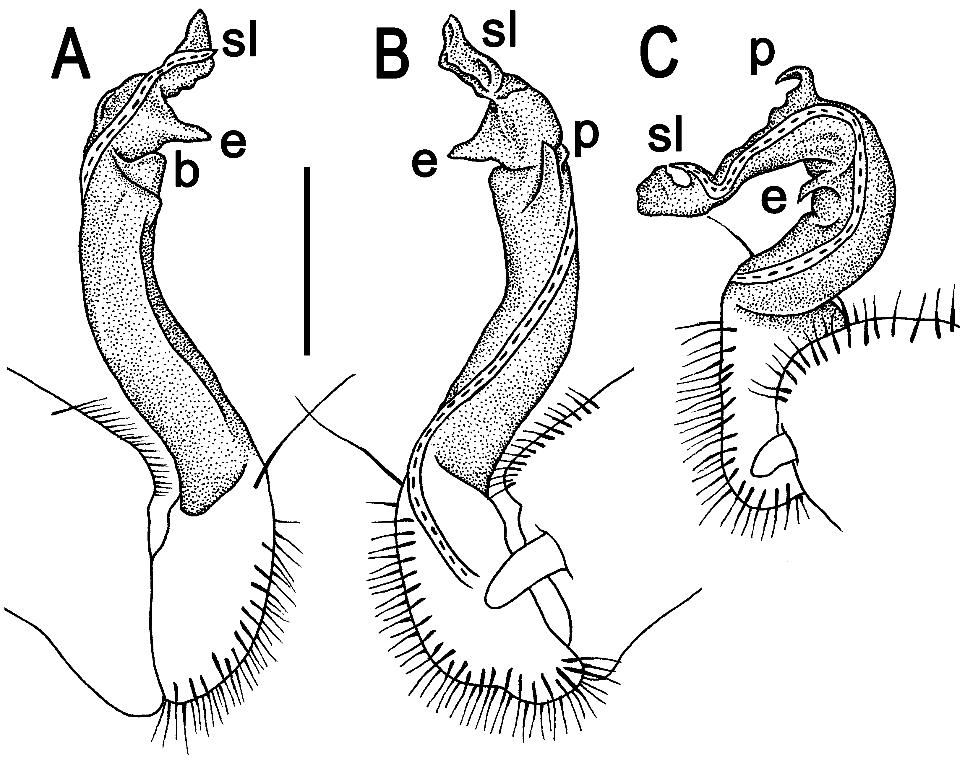
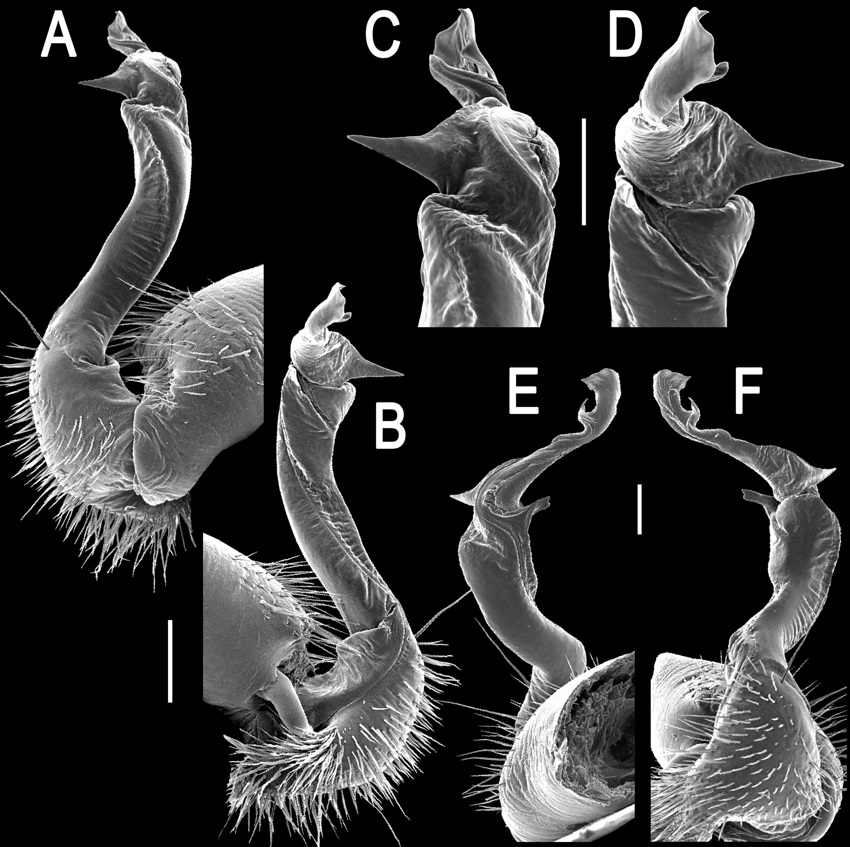
Gonopods ( Figs 9 View FIGURE 9 & 10 View FIGURE 10 ) with large, long, distoventrally sparsely setose coxae. Femorite about 2 times as long as prefemoral part. Femorite rather stout, long and curved; seminal groove running mesally along entire femorite, with an evident apicoventral shelf ( b) and a pointed, parabasal subventral process p; process e prominent, spearshaped, short and pointed; “postfemoral” portion demarcated by a distinct lateral sulcus. Solenomere ( sl) strong, twisted, with a short subapical branchlet terminating seminal groove.

| CUMZ |
Chulalongkorn University Museum of Natural History |
No known copyright restrictions apply. See Agosti, D., Egloff, W., 2009. Taxonomic information exchange and copyright: the Plazi approach. BMC Research Notes 2009, 2:53 for further explanation.
|
Kingdom |
|
|
Phylum |
|
|
Class |
|
|
Order |
|
|
Family |
|
|
Genus |