
Tuberculobasis
|
publication ID |
https://doi.org/10.5281/zenodo.187806 |
|
DOI |
https://doi.org/10.5281/zenodo.4391363 |
|
persistent identifier |
https://treatment.plazi.org/id/1312774B-FFD7-FFE4-FF04-F911FA3BFE69 |
|
treatment provided by |
Plazi |
|
scientific name |
Tuberculobasis |
| status |
|
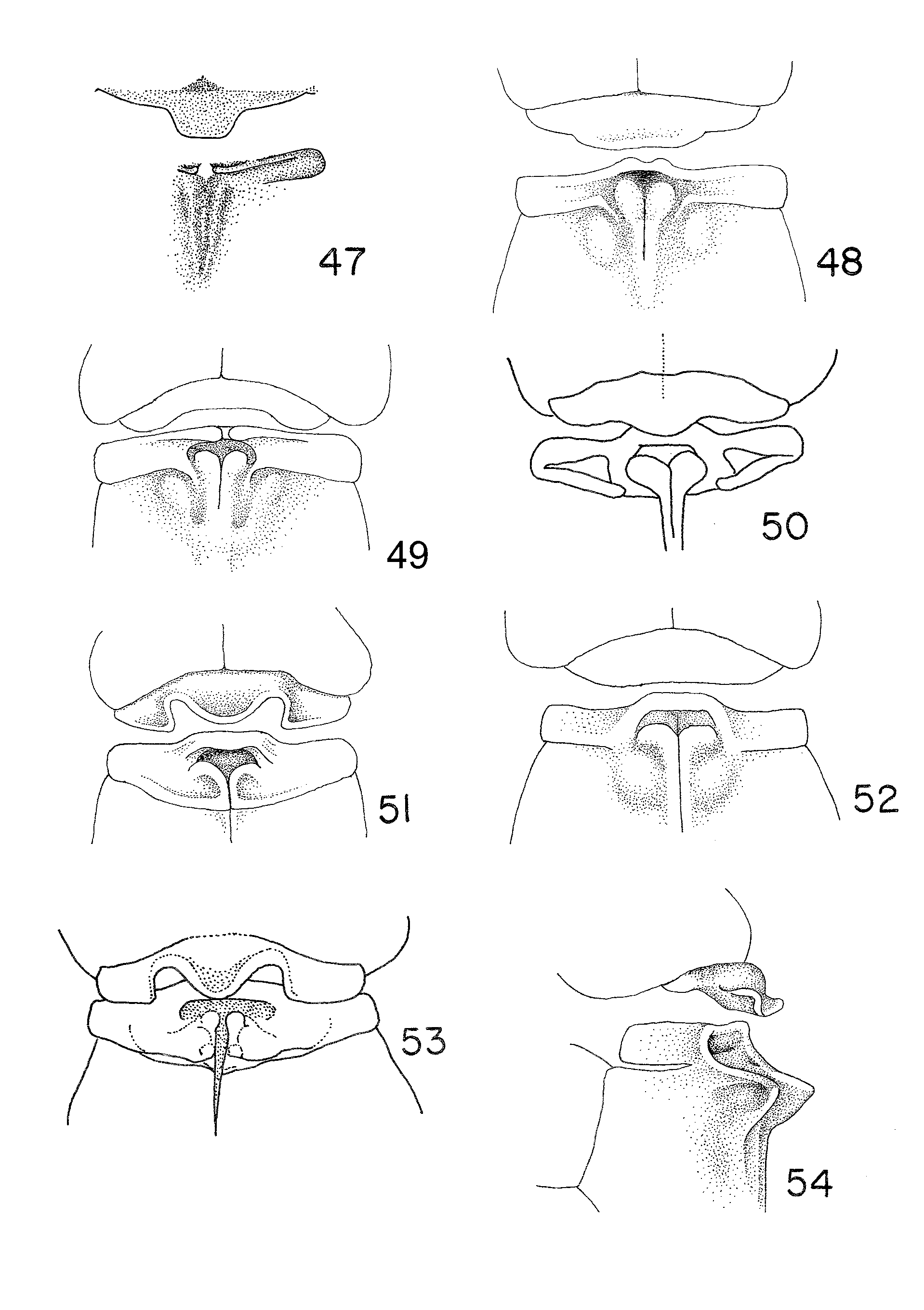
Key to females of Tuberculobasis View in CoL
(Females of T. arara , T. geijskesi , T. guarani , T. karitiana , and T. macuxi unknown)
1. With mesepisternal tubercles ( Figs 48, 49, 52 View FIGURES 47 – 54 )............................................................................................................. 2
1´. Without mesepisternal tubercles ( Figs 50, 51, 53 View FIGURES 47 – 54 ) ....................................................................................................... 5
2. Mesepisternal tubercles not visible in dorsal view ( Fig. 47 View FIGURES 47 – 54 ) and scarcely visible in dorso-lateral view. Vulvar spine of S8 absent ( Fig. 95 View FIGURES 95 – 100 )................................................................................................................................. T. cardinalis View in CoL
2´. Mesepisternal tubercles visible in dorsal view ( Figs 48, 49, 52 View FIGURES 47 – 54 ). Vulvar spine of S8 present ( Figs 96–100 View FIGURES 95 – 100 ).............. 3
3. Mesepisternal tubercles connected with anterior margin of mesostigmal plate ( Fig. 52 View FIGURES 47 – 54 ) ....................... T. williamsoni View in CoL
3´. Mesepisternal tubercles not connected with the anterior margin of mesostigmal plates ( Figs 48, 49 View FIGURES 47 – 54 ) ....................... 4
4. Median lobe of hind prothoracic lobe depressed and poorly defined ( Fig. 49 View FIGURES 47 – 54 ).............................................. T. inversa View in CoL
4´. Median lobe of hind prothoracic lobe elevated and well defined ( Fig. 48 View FIGURES 47 – 54 ).............................................. T. costalimai View in CoL
5. Median lobe of hind prothoracic lobe separated from lateral lobes by two deep concavities ( Figs 51, 53 View FIGURES 47 – 54 ). ............... 6
5´. Median lobe of hind prothoracic lobe separated from lateral lobes by two shallow concavities ( Fig. 50 View FIGURES 47 – 54 )..................................................................................................................................................................................... T. mammilaris View in CoL
6. Hind margin of mesostigmal plate curved anteriorly and laterally to form a C-shaped structure ( Fig. 51 View FIGURES 47 – 54 )......... T.tirio View in CoL
6´. Hind margin of mesostigmal plate not curved to form a C-shaped structure ( Fig. 53 View FIGURES 47 – 54 ) .............................. T.yanomami View in CoL

No known copyright restrictions apply. See Agosti, D., Egloff, W., 2009. Taxonomic information exchange and copyright: the Plazi approach. BMC Research Notes 2009, 2:53 for further explanation.
|
Kingdom |
|
|
Phylum |
|
|
Class |
|
|
Order |
|
|
Family |
|
|
Genus |